Glutes

The muscles in the group are:

Gluteus Maximus

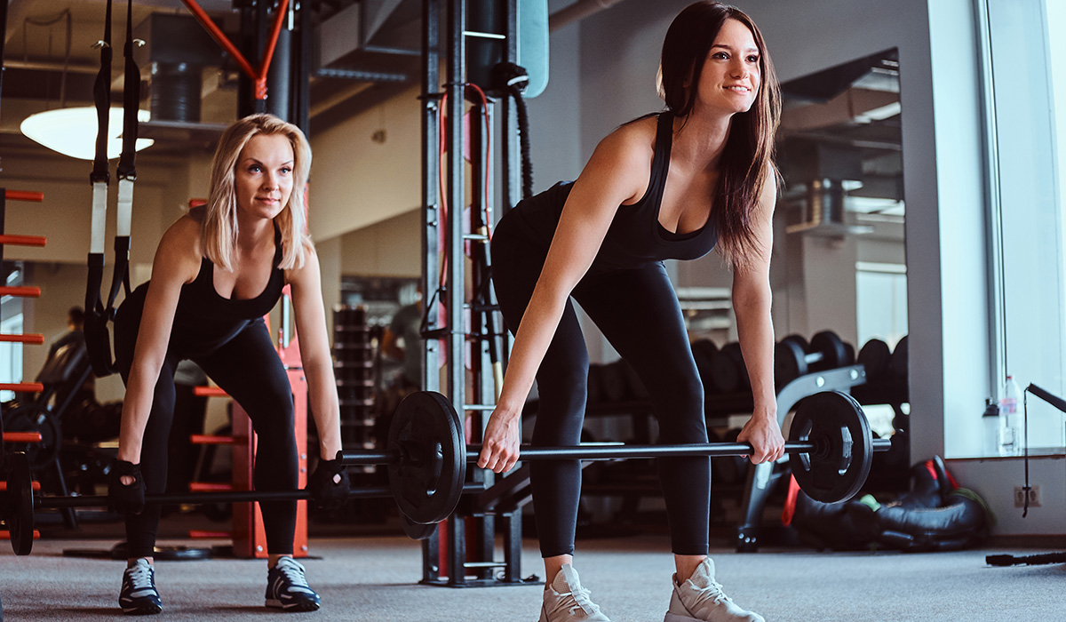
Primary function is upper leg (thigh) extension. (i.e., moving the upper leg backwards as in rising from a squat position). The same with bent-leg deadlifting, the rear leg drive when sprinting and any hip extension exercise where the thigh is extended backwards.

Gluteus Medius and Minimus

Perform similar functions depending on the position of the knee and hip joints. With the knee extended, they abduct the thigh (out to the side away from the opposite leg). When running, they stabilize the leg during the single-support phase. With the hips flexed, they internally rotate the thigh. With the hips extended, they externally rotate the thigh.

Gamellus Inferior and Superior

Both assist to laterally rotate the extended thigh.

Quadratus Femoris

A deep muscle in your gluteal region and is generally concerned with lateral rotation and stabilisation of the femur at the hip joint and is a strong external rotator and adductor of the thigh.

Obturator Externus

Primary action is to externally rotate the femur when the hip was in neutral position and flexed at 90°. Its secondary function is as an adductor when the hip was in flexion.

Piriformis

Helps rotate the hip and works with rotators such as the obturator externus and the gemellus inferior. It will rotate the thigh while extended and will abduct, or pull inward, the thigh when flexed.
Triceps

Triceps brachii comes from the Latin for “three-headed muscle of the arm”.

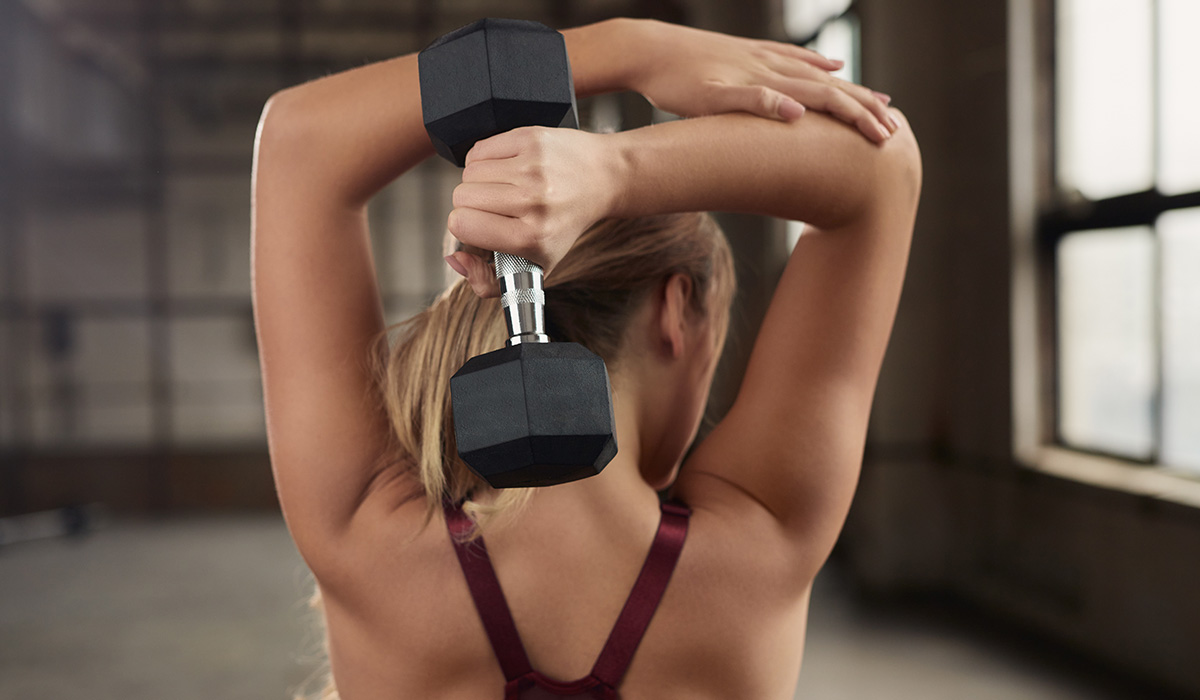
It is the large muscle on the back of your arms and is responsible for extension of the elbow. It is also the antagonist to the biceps brachii and brachialis muscles. The triceps help to fixate the elbow when the hand is involved in fine or intricate movements such as writing. It originates from the scapula, so therefore also becomes a stabilising muscle for the shoulder joint and aids adduction of the arm. The muscle is also capable of contracting statically.

The triceps muscles can be trained either through isolation or compound extension movements. Examples of isolation movements would include cable push downs and extensions where the arms go behind the back.
Examples of compound movements include close grip bench press and military press. A close grip on the bar really targets the triceps more than a wider grip.

You should always train your triceps through their full range of movement as they cross two joints (shoulder and elbow).

It is very rare to rupture or tear your triceps muscles.

Calves

The calf muscle, on the back of the lower leg, is made up of two muscles:

THE GASTROCNEMIUS
This is the larger calf muscle, forming the bulge visible beneath the skin. The gastrocnemius has two parts or “heads,” which together create its diamond shape.

THE SOLEUS
This is a smaller, flat muscle that lies underneath the gastrocnemius muscle. The gastrocnemius and soleus muscles taper and merge at the base of the calf muscle. Tough connective tissue at the bottom of the calf muscle merges with the Achilles tendon. The Achilles tendon inserts into the heel bone (calcaneus). During walking, running, or jumping, the calf muscle pulls the heel up to allow forward movement.

Hamstrings

BICEPS FEMORIS – Not to be confused with the bicep muscle in your upper arm!! Its main action is flexion at the knee, but it is also responsible for extending the thigh at the hip and lateral rotation at the hip and knee.

SEMITENDINOSUS – Responsible for flexion of the leg at the knee joint and extension of thigh at the hip. It also medially rotates the thigh at the hip joint and the leg at the knee joint.

SEMIMEMBRANOSUS – Lies underneath the semitendinosus. It is responsible for flexion of the leg at the knee joint, extension of the thigh at the hip and medial rotation of the thigh at the hip joint and the leg at the knee joint.

If you spend much of your day sat behind a desk, then it is not unusual for your hamstrings to feel tight – however, it might not solely be your hamstrings causing the issue. If your hip flexors and the muscles in the front of your pelvis are tight, this can elevate the attachments of your hamstrings (it causes an anterior tilt in the pelvis and basically places your hamstrings in a lengthened position) which in turn creates that feeling of tightness. Make sure you stretch your hip flexors out as well as your hamstrings before and after exercise!

Shoulders

THE DELTOIDS
‘Delts’ are teardrop shaped and have three parts, anterior, medial and posterior. They control the flexion, abduction and extension of the humerus.

ROTATOR CUFF
Made up of four muscles (see below) – the supraspinatus, infraspinatus, teres minor and subscapularis. These help to stabilise the shoulder joint and assist with the abduction, adduction and rotation of the humerus. Due to their location, they are prone to small tears and impingements.

Supraspinatus – This helps to hold the humerus in place and to lift the arm.
Infraspinatus – This is the main muscle for rotation and extension of the shoulder.
Teres minor – This is the smallest of the rotator cuff muscles and its main role is to help with the rotation of the arm away from the body.
Subscapularis – This is what holds the humerus to the shoulder blade and aids the rotation of the arm and allows you to hold your arm straight out and to lower it.

LEVATOR SCAPULAE
This helps with the elevation of the shoulder blade.

TRAPEZIUS
These allow your scapula to elevate, retract and depress.

RHOMBOIDS
These can be found under your trapezium and help with the elevation and retraction of the scapula.

Chest

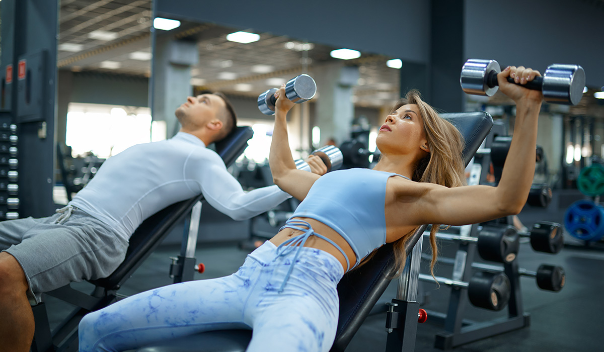
The chest forms part of a larger group of “pushing” muscles in the upper body and is made up of three muscles: pectoralis major, minor and the serratus anterior.

PECTORALIS MAJOR

This is the larger of the chest muscles and is responsible for adduction of the arms, rotation of the arm forward and when the arms are raised in a fixed position (like you are wall climbing) it works with the teres major and the latissimus dorsi to pull your torso upwards.

PECTORALIS MINOR

This muscle sits below the pectoralis major and it attaches into the scapula. It is responsible for pulling the shoulder forward and down.

SERRATUS ANTERIOR

Also known as the “boxer’s muscle”. Although it might not be a true chest muscle, it is classed in this group because of its attachment on the ribs near the pectoral muscles.

Abductors and Adductors

Abductors

This group of muscles are responsible for hip abduction (moving the leg away from the midline of the body).

They also help with rotation of the leg at the hip joint and are necessary for being able to remain stable when walking or standing on one leg. In many people, these muscles tend to be weak which leads to walking and posture issues.

Other muscles included in the abduction movement are the sartorius, piriformis, glute maximus and the ITB.

When these muscles are weak, it can lead to injuries such as Achilles’ tendinopathy, patellofemoral pain syndrome or runner’s knee, IT band syndrome, hamstring tendinopathy or plantar fasciitis.

Strength training can work well to reduce pain and strong muscles and tissues can protect against injury.

If you are injured then strength training will help heal you.

Adductors

An adductor muscle is any of the muscles that draws /pulls a part of your body towards the median line or towards the axis of an extremity. (You have adductor muscles in your thumb and big toe!)

The three powerful muscles in our thighs that make up the adductor group are the adductor longus, adductor brevis and the adductor magnus. They are ribbon like and are attached along the femur. Their primary action is adduction of the thigh, like squeezing your thighs together and they also help in the rotation and flexion of the hip.

The pectineus and gracilis, are also part of the adductor group of muscles.

If you experience “groin pull/strain” it will usually be one of these muscles that is causing it.

Other ailments related to imbalanced adductors include arthritis (ankle, knee, hip), plantar fasciitis, ankle sprain, chondromalacia (damage to the cartilage in the knee), IT band strain, piriformis syndrome, sciatica, and low back pain.

Biceps

The biceps muscle has 2 heads, the long head and short head.

BICEPS BRACHII

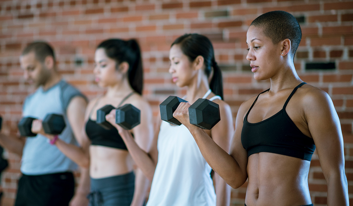
This muscle is responsible for bending the forearm back towards the upper arm so is involved in lifting and pulling movements. It also plays a part in supinating the forearm, turning the palms to face upwards or forwards.

The biceps muscles lie between the shoulder and the elbow. It is one of three muscles that make up the front (or anterior) part of the upper arm. It shares the space with the Brachialis muscle and the Coracobrachialis muscle.

The biceps muscle crosses the shoulder and the elbow, making it one of the few muscles that cross more than one joint.

BRACHIALIS

This smaller muscle is located underneath the biceps brachii and on both sides. The brachialis assists with flexion of the elbow. One unique fact about this muscle is that it only becomes completely activated when the arm is being flexed but not actually moving.

Core

The core transfers the forces between your upper and lower extremities. The core and limbs need to be strong and stable before any form of movement can happen through the limbs so, the stronger you make your core, the more efficient and stronger your movements will be.

Your core comprises of more than just your abs and can be split into two categories: stabilisers and movers. Think of your core muscles as any muscle that are attached to the spine.

STABILISERS

  • Transverse Abominais
  • Internal Obliques
  • Lumbar
  • Multifidus Pelvic floor muscles
  • Diaphragm
  • Transverse spinalis

MOVERS

  • Rectus Abdominis
  • External Obliques
  • Erector Spinae
  • Latissimus Dorsi
  • Hamstrings
  • Hip abductors and adductors

Upper Back

The muscles in the group are:

TRAPEZIUS

This is broad, flat and triangular in shape. It is responsible for elevating and retracting the scapula (shoulder blade) and when the arm abducts (moves away from the body’s centre line), it rotates the scapula.

LATISSIMUS DORSI

The latissimus dorsi muscles, known as the lats, are the large V-shaped muscles that connect your arms to your vertebral column. They help protect and stabilize your spine while providing shoulder and back strength. Your lats also help with shoulder and arm movement and support good posture.

LEVATOR SCAPULAE

This is a smaller muscle attached between the neck and scapula. Its main function is to elevate the scapula.

RHOMBOIDS

As shown in the diagram, there are two muscles, major and minor. Both muscles are responsible for the retraction and rotation of the scapula.